 ALERT
ALERT ATTENTION ⚠️
In observance of a holiday, Agilent CrossLab/iLab Operations Software Support Help Desk will be closed during U.S. hours on Friday, July 3rd, 2026. We will resume regular U.S. support hours on Monday, July 6th, 2026. EU and APAC Support will remain open during this time. For urgent matters, please add "Urgent" to the ticket/email subject or press "1" when prompted to escalate a call on the iLab Support phone, and we will prioritize those requests first.
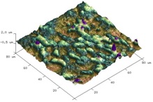

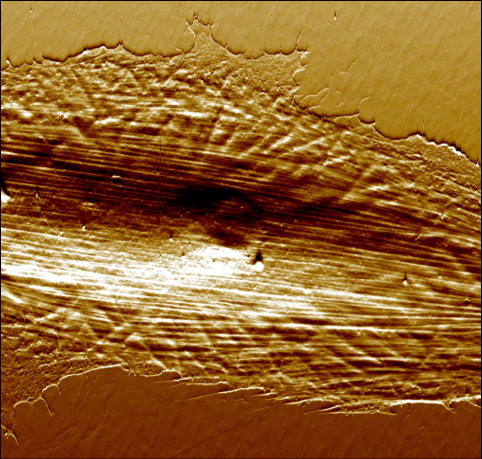
Atomic Force Microscopy (AFM) is a noninvasive technique that produces high-resolution topographic images under physiological-like conditions. Scanning can be performed in air or in a liquid environment, allowing imaging of a wide range of samples, from living cells down to single molecules.
AFM is also a powerful tool for studying dynamic processes such as cellular endocytosis of nanovectors and systemic responses to biological events. A major area of interest is determining sample stiffness (Elastic modulus) to assess experimental data. The elasticity of the sample surface can vary between cell/tissue types and change as a function of growth, differentiation, disease, or treatment. In addition, AFM can image non-cellular structures with exceptional detail.
With the recent acquisition of our NanoWizard V AFM (NIH grant 1S10OD036249), we can now perform PeakForce Quantitative Imaging (PFQI), enabling precise measurement of mechanical properties such as stiffness, adhesion, and modulus at every pixel while capturing high-resolution topography.
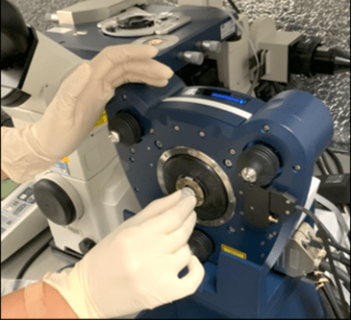
The core utilizes a NanoWizard V Atomic Force Microscope (JPK, Bruker Nano Inc.) that requires minimal sample preparation. This system is integrated with a Nikon TE2000 inverted optical microscope, enabling simultaneous acquisition of bright-field and fluorescence images of samples under study.
We welcome inquiries from internal, external, and public sector partners. Please contact us for more information.
|
Ana Maria Zaske, PhD |
|
This work was supported by the NIH Shared Instrumentation Grant S10OD036249‑01A1, which funded the NanoWizard V Atomic Force Microscope used in this study at the AFM Core Facility, McGovern Medical School, UTHealth Houston.
| Hours | Location |
|
Monday - Friday 9 am - 5 pm |
Internal Medicine, UT-Health |
| Name | Role | Phone | Location | |
|---|---|---|---|---|
| Ana Maria Zaske, PhD |
Director
|
713-486-5418
|
Ana.M.Zaske@uth.tmc.edu
|
1881 EAST RD, 3SCRB6.3728, HOUSTON, TX 77054
|
